NEWS
Non-averaged single-molecule 3D structure determined by individual-particle cryo-electron tomography (IPET)
Life is in motion, cells are in motion, atoms undergo thermal vibration—how could macromolecular structures not be in motion? Despite knowing that macromolecular particles experience Brownian motion in solution, with reaction forces from solvent atoms causing vibrations, the macromolecular structures determined by current experimental techniques are presented as static.
This is because current structural techniques, such as X-ray crystallography and cryo-electron microscopy (cryo-EM) single-particle averaging (SPA), rely on averaging methods. By this process, a small, homogeneous subset of particles is selected from a heterogeneous population and averaged to produce a static 3D structure. While this improves the signal-to-noise ratio (SNR) and enables high-resolution reconstruction of rigid domains, it often results in the loss of density in flexible regions and introduces anisotropic resolution in the final 3D map. As a result, the structural variability of the majority of unselected particles remains unexplored, limiting our ability to capture macromolecular flexibility.
Furthermore, resolving low-resolution 3D structures of macromolecules with intrinsic structural flexibility and continuous conformational changes remains a major challenge. Thus, single-molecule 3D structure determination, without the need for averaging, is a highly sought-after approach to understanding macromolecular dynamics and mechanisms during chemical reactions.
The objective of this laboratory is to develop a cryo-electron tomography (cryo-ET) method to determine the 3D structure of a single molecule without averaging, named the individual-particle electron tomography (IPET) method (Zhang, et al., PLoS One, 2012), without averaging. Structure determination of a single molecule plays an important role in understanding macromolecular dynamics and the function/mechanism during chemical reactions. However, this determination is not trivial, but rather challenging due to the extremely low signal-to-noise ratio (SNR) of a single molecule caused by the electron dose limit necessary for preventing radiation damage. Over the past decade, this laboratory has reported a series of TEM techniques to improve the TEM capability to image a single molecule.
The IPET related TEM techniques have been reported include:
i) The development of focused 3D reconstruction algorithms to reduce the large-scale image distortion/deformation effects on 3D reconstruction (Zhang, et al., PLoS One, 2012);
ii) A fully mechanical-controlled TEM software for cryo-ET data acquisition to prevent beam-hysteresis caused by beam-coherence reduction during tilting (Liu, et al., Sci Rep., 2016);
iii) A noise-reducing algorithm to enhance the signal-to-noise ratio (SNR) of a single molecule (Wu, et al., Sci Rep., 2018);
iv) A missing-wedge correction method to reduce the enlargement artifact induced by the tilt limit (Zhai, et al., Sci Rep., 2020).
Other TEM methods were also reported as following:
v) A cryo-positive staining (cryo-PS) method for imaging the detailed structure of a single-instant small protein (Zhang, et al., Nat Chem Bio., 2012);
vi) An optimized negative-staining (OpNS) method to reduce the relaxation artifact of lipoproteins (Rames, et al., J Vis Exp., 2014; Zhang, et al., J Lipid Res., 2010);
vii) A simple liquid-cell TEM method for real-time imaging the dynamics of liquid-vapor interface (Ren, et al., Sci Rep., 2020).
Based on current improvements, this laboratory has characterized the structural variety of several flexible macromolecules, including:
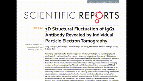
1) Antibodies (Zhang, et al., Sci Rep., 2015; Tong, et al., Sci Rep., 2013; Zhang, et al. Anal Chem., 2017; Lei, at al., Sci Rep., 2020),
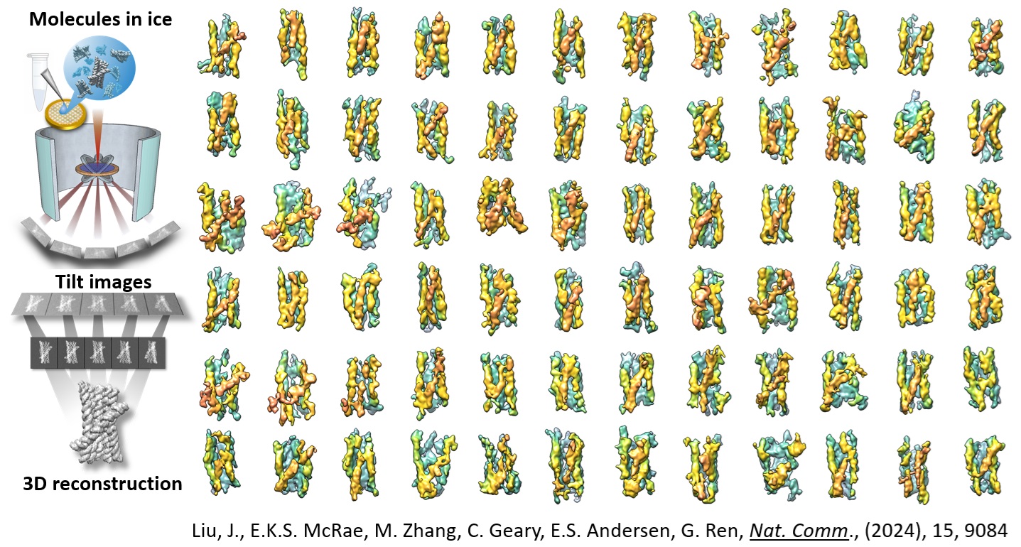
2) D/RNA-related macromolecules (Zhang, et al., Nat. Comm., 2016; Lei, et al., Nat. Comm., 2021; Wang, et al., Nat Comm., 2022; Zhang, et al., Mol. Cell., 2022; McRae, et al., Nat Nano., 2023; Zhang, et al., Nat. Comm, 2022; Liu, et al., Nat. Comm., 2023),
3) Lipoproteins (Zhang, et al., Sci Rep., 2015; Yu, et al.,J Lipid Res., 2016; Lei, et al.,BBA lipids, 2019),
4) Neuron proteins (Lu, et al., J Bio Chem., 2014; Lu, et al., J Biol Chem., 2016; Lee, et al., J Biol Chem., 2023).
5) Nanoparticles (Xu, et al., Nat. Energy., 2023; Xie, et al., Nanoscale Advances, 2023).
The development of IPET capability for high-resolution structure determination of a single protein is crucial in translating the current static structures into future dynamic structures, which is essential for understanding the processes of synthesis, folding, and chemical reactions of macromolecules.